We wouldn`t want to lose sight of the big picture because of a hiccup or two along the tortuous route to regulatory approval, IT WILL HAPPEN because it needs to happen!!!
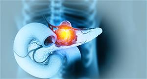
The authors evaluated the feasibility of using the first clinical-grade confocal laser endomicroscopy (CLE) system using fluorescein sodium for intraoperative in vivo imaging of brain tumors.METHODS A CLE system cleared by the FDA was used in 30 prospectively enrolled patients with 31 brain tumors (13 gliomas, 5 meningiomas, 6 other primary tumors, 3 metastases, and 4 reactive brain tissue). A neuropathologist classified CLE images as interpretable or noninterpretable. Images were compared with corresponding frozen and permanent histology sections, with image correlation to biopsy location using neuronavigation. The specificities and sensitivities of CLE images and frozen sections were calculated using permanent histological sections as the standard for comparison. A recently developed surgical telepathology software platform was used in 11 cases to provide real-time intraoperative consultation with a neuropathologist.
RESULTS Overall, 10,713 CLE images from 335 regions of interest were acquired. The mean duration of the use of the CLE system was 7 minutes (range 3–18 minutes). Interpretable CLE images were obtained in all cases. The first interpretable image was acquired within a mean of 6 (SD 10) images and within the first 5 (SD 13) seconds of imaging; 4896 images (46%) were interpretable. Interpretable image acquisition was positively correlated with study progression, number of cases per surgeon, cumulative length of CLE time, and CLE time per case (p ≤ 0.01). The diagnostic accuracy, sensitivity, and specificity of CLE compared with frozen sections were 94%, 94%, and 100%, respectively, and the diagnostic accuracy, sensitivity, and specificity of CLE compared with permanent histological sections were 92%, 90%, and 94%, respectively. No difference was observed between lesion types for the time to first interpretable image (p = 0.35). Deeply located lesions were associated with a higher percentage of interpretable images than superficial lesions (p = 0.02). The study met the primary end points, confirming the safety and feasibility and acquisition of noninvasive digital biopsies in all cases. The study met the secondary end points for the duration of CLE use necessary to obtain interpretable images. A neuropathologist could interpret the CLE images in 29 (97%) of 30 cases.
CONCLUSIONS The clinical-grade CLE system allows in vivo, intraoperative, high-resolution cellular visualization of tissue microstructure and identification of lesional tissue patterns in real time, without the need for tissue preparation.
https://www.neurosurgery-blog.com/archives/author/admin
We wouldn`t want to lose sight of the big picture because of a...


Add OIL (ASX) to my watchlist
 (20min delay) (20min delay)
|
|||||
|
Last
20.5¢ |
Change
0.000(0.00%) |
Mkt cap ! $171.2M | |||
| Open | High | Low | Value | Volume |
| 20.5¢ | 20.5¢ | 20.0¢ | $15.10K | 75.16K |
Buyers (Bids)
| No. | Vol. | Price($) |
|---|---|---|
| 1 | 292 | 20.5¢ |
Sellers (Offers)
| Price($) | Vol. | No. |
|---|---|---|
| 21.0¢ | 16440 | 2 |
View Market Depth
| Last trade - 16.10pm 15/08/2024 (20 minute delay) ? |
| OIL (ASX) Chart |