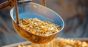
For interest, some images from a case. Patient with metastatic colorectal cancer received SIRT and standard chemo, excellent response allowed for resection of the liver metastasis. SirSpheres are the round purple beads centre field. Top left is normal liver, bottom right shows broad zones of dead tumour, there were only very rare microscopic foci of cancer left (linear purple glands, centre of bottom image).
Can't assess unresected tumours in this way (PET for metabolic activity is best alternative), but with this degree of tumour kill it would be unlikely that the minimal tumour would be the site of first progression. First liver progression in the SIRT arm of SIRFLOX was more likely to occur at a site other than the treated lesion, easy to see why.


For interest, some images from a case. Patient with metastatic...


Add to My Watchlist
What is My Watchlist?